-->

Wednesday, February 18, 2009

Beauty Tips

http://www.justimaging.co.za/samples/beauty/images/Banner.jpg

Featured Articles:


Laser Hair Removal - Effectively remove unwanted hair
Get Rid of Acne Scars - Reduce the appearance of acne scars.
Versatile Haircuts - All-around haircuts for any occasion
Natural Moisturizers for Dry Skin - Secrets for staying soft
Natural Tips for Shiny Hair - Head to the kitchen!
Natural Ways To Lighten Your Skin - Dark spots, and freckles, and skin-damage - oh my!
Sexy Women - How to look sexy without being sleazy

https://blogger.googleusercontent.com/img/b/R29vZ2xl/AVvXsEiaqeORZRcfNqnFSxt-py2sRH-awlphADCdxtyrFny3-gBxl8gUr888TdpvP-AhCSXwKNAFB-s3s-19JRzA5YgYI87o29ieQN1lM8efser2znk-JUV3mHvwUCknK4Hg3eeHiv0QHaiBzyxM/s1600/coolbeautytips_logo.gif

Monday, February 16, 2009

Cardiac arrhythmia

Cardiac arrhythmia (also dysrhythmia) is a term for any of a large and heterogeneous group of conditions in which there is abnormal electrical activity in the heart. The heart beat may be too fast or too slow, and may be regular or irregular.

Some arrhythmias are life-threatening medical emergencies that can result in cardiac arrest and sudden death. Others cause aggravating symptoms such as an abnormal awareness of heart beat (palpitations), and may be merely annoying. Others may not be associated with any symptoms at all, but pre-dispose toward potentially life threatening stroke or embolus.

Some arrhythmias are very minor and can be regarded as normal variants. In fact, most people will sometimes feel their heart skip a beat, or give an occasional extra strong beat - neither of which is usually a cause for alarm.

The term sinus arrhythmia refers to a normal phenomenon of mild acceleration and slowing of the heart rate that occurs with breathing in and out. It is usually quite pronounced in children, and steadily lessens with age. This can also present during meditation breathing exercises that involve deep inhaling and breath holding patterns.

Manifestations

The term cardiac arrhythmia covers a very large number of very different conditions.

The most common symptom of arrhythmia is an abnormal awareness of heartbeat, termed palpitations. These may be infrequent, frequent, or continuous. Some of these arrhythmias are harmless (though annoying) but many of them predispose to adverse outcomes.

Some arrhythmias do not cause symptoms, and are not associated with increased mortality. However, some asymptomatic arrhythmias are associated with adverse events. Examples include increase in risk of blood clotting within the heart, and also an insufficient amount of blood is transported to the heart because of weak heart beat, and thus increase the risk of embolisation and stroke, or increase in the risk of heart failure, or increase in the risk of sudden cardiac death.

If an arrhythmia results in a heart beat that is too fast, too slow or too weak to supply the body's needs, this manifests as a lower blood pressure and may cause lightheadedness or dizziness, or fainting.

Some types of arrhythmia result in cardiac arrest, or sudden death.

Medical assessment of the abnormality using an electrocardiogram is the best way to diagnose and assess the risk of any given arrhythmia.

Mechanisms and etiology

Normal electrical activity in the heart

Each heart beat originates as an electrical impulse from a small area of tissue in the right atrium of the heart called the sinus node or Sino-atrial node or SA node. The impulse initially causes both of the atria to contract, then activates the atrioventricular (or AV) node which is normally the only electrical connection between the atria and the ventricles or main pumping chambers. The impulse then spreads through both ventricles via the His Purkinje fibres causing a synchronised contraction of the heart muscle, and thus, the pulse.

In adults the normal resting heart rate ranges from 60 to 100 beats per minute. The resting heart rate in children is much faster.

Bradycardias

Normal sinus rhythm, with solid black arrows pointing to normal P waves representative of normal sinus node function, followed by a pause in sinus node activity (resulting in a transient loss of heart beats). Note that the P wave that disrupts the pause (indicated by the dashed arrow) does not look like the previous (normal) P waves- this last P wave is arising from a different part of the atrium, representing an escape rhythm.

A slow rhythm, (less than 60 beats/min), is labelled bradycardia. This may be caused by a slowed signal from the sinus node (termed sinus bradycardia), a pause in the normal activity of the sinus node (termed sinus arrest), or by blocking of the electrical impulse on its way from the atria to the ventricles (termed AV block or heart block). Heart block comes in varying degrees and severity. It may be caused by reversible poisoning of the AV node (with drugs that impair conduction) or by irreversible damage to the node.

Tachycardias

Any heart rate faster than 100 beats/minute is labelled tachycardia. Tachycardia may result in palpitation, however, tachycardia is not necessarily an arrhythmia. Increased heart rate is a normal response to physical exercise or emotional stress. This is mediated by the sympathetic nervous system on the sinus node, and is called sinus tachycardia. Other things that increase sympathetic nervous system activity in the heart include ingested or injected substances such as caffeine or amphetamines, and an overactive thyroid gland (hyperthyroidism).

Tachycardia that is not sinus tachycardia usually results from the addition of abnormal impulses to the normal cardiac cycle. Abnormal impulses can begin by one of three mechanisms: automaticity, reentry or triggered activity. A specialised form of re-entry problem is termed fibrillation.

Automaticity

Automaticity refers to a cardiac muscle cell firing off an impulse on its own. All of the cells in the heart have the ability to initiate an action potential, however, only some of these cells are designed to routinely trigger heart beats. These cells are found in the 'conduction system' of the heart and include the SA node, AV node, Bundle of HIS and Purkinje fibers. The sinoatrial node is a single specialized location in the atrium which has a higher automaticity (a faster pacemaker) than the rest of the heart, and therefore is usually responsible for setting the heart rate, and initiating each heart beat.

Any part of the heart that initiates an impulse without waiting for the sinoatrial node is called an ectopic focus, and is by definition a pathological phenomenon. This may cause a single premature beat now and then, or, if the ectopic focus fires more often than the sinoatrial node, it can produce a sustained abnormal rhythm. Rhythms produced by an ectopic focus in the atria, or by the atrioventricular node, are the least dangerous dysrhythmias; but they can still produce a decrease in the heart's pumping efficiency, because the signal reaches the various parts of the heart muscle with different timing to usual and can be responsible for poorly coordinated contraction.

Conditions that increase automaticity include sympathetic nervous system stimulation and hypoxia. The resulting heart rhythm depends on where the first signal begins: if it is the sinoatrial node, the rhythm remains normal but rapid; if it is an ectopic focus, many types of dysrhythmia can result.

Re-entry

Re-entry arrhythmias occur when an electrical impulse recurrently travels in a tight circle within the heart, rather than moving from one end of the heart to the other and then stopping. Every cardiac cell is able to transmit impulses in every direction, but will only do so once within a short period of time. Normally, the action potential impulse will spread through the heart quickly enough that each cell will only respond once. However, if conduction is abnormally slow in some areas, part of the impulse will arrive late and potentially be treated as a new impulse. Depending on the timing, this can produce a sustained abnormal circuit rhythm. Re-entry circuits are responsible for atrial flutter, most paroxysmal supraventricular tachycardia, and dangerous ventricular tachycardia.

By analogy, imagine a room full of people all given these instructions: "If you see anyone starting to stand up, then stand up for three seconds and sit back down." If people are quick enough to respond, the first person to stand will trigger a single wave which will then die out; but if there are stragglers on one side of the room, people who have already sat down will see them and start a second wave, and so on.

Fibrillation

When an entire chamber of the heart is involved in a multiple micro-reentry circuits, and therefore quivering with chaotic electrical impulses, it is said to be in fibrillation.

Fibrillation can affect the atrium (atrial fibrillation) or the ventricle (ventricular fibrillation); ventricular fibrillation is imminently life-threatening.

Atrial fibrillation affects the upper chambers of the heart, known as the atria. Atrial fibrillation may be due to serious underlying medical conditions, and should be evaluated by a physician. It is not typically a medical emergency.

Ventricular fibrillation occurs in the ventricles (lower chambers) of the heart; it is always a medical emergency. If left untreated, ventricular fibrillation (VF, or V-fib) can lead to death within minutes. When a heart goes into V-fib, effective pumping of the blood stops. V-fib is considered a form of cardiac arrest, and an individual suffering from it will not survive unless cardiopulmonary resuscitation (CPR) and defibrillation are provided immediately.

CPR can prolong the survival of the brain in the lack of a normal pulse, but defibrillation is the only intervention which can restore a healthy heart rhythm. Defibrillation is performed by applying an electric shock to the heart, which resets the cells, permitting a normal beat to re-establish itself.

Triggered beats

Triggered beats occur when problems at the level of the ion channels in individual heart cells result in abnormal propagation of electrical activity and can lead to sustained abnormal rhythm. They are relatively rare, but can result from the action of anti-arrhythmic drugs.

Classification of common cardiac arrhythmias

Arrhythmia may be classified by rate (normal, tachycardia, bradycardia), or mechanism (automaticity, reentry, fibrillation).

It is also appropriate to classify by site of origin:

Atrial

  • Premature Atrial Contractions (PACs)
  • Wandering Atrial Pacemaker
  • Multifocal atrial tachycardia
  • Atrial flutter
  • Atrial fibrillation (Afib)

Junctional arrhythmias

  • Supraventricular tachycardia (SVT)
  • AV nodal reentrant tachycardia is the most common cause of Paroxysmal Supra-ventricular Tachycardia (PSVT)
  • Junctional rhythm
  • Junctional tachycardia
  • Premature junctional complex

Atrio-ventricular

  • AV reentrant tachycardia occurs when a re-entry circuit crosses between the atria and ventricles somewhere other than the AV node:
    • Wolff-Parkinson-White syndrome
    • Lown-Ganong-Levine syndrome

Ventricular

  • Premature Ventricular Contractions (PVC) sometimes called Ventricular Extra Beats (VEBs)
    • Premature Ventricular beats occurring after every normal beat are termed ventricular bigeminy
    • Two premature ventricular beats for each normal beat is termed ventricular trigeminy
    • Three premature ventricular beats for each normal beat is termed "A Run of V-Tach"
  • Accelerated idioventricular rhythm
  • Monomorphic Ventricular tachycardia
  • Polymorphic ventricular tachycardia
  • Ventricular fibrillation

Heart blocks

These are also known as AV blocks, because the vast majority of them arise from pathology at the atrioventricular node. They are the most common causes of bradycardia:

  • First degree heart block, which manifests as PR prolongation
  • Second degree heart block
    • Type 1 Second degree heart block, also known as Mobitz I or Wenckebach
    • Type 2 Second degree heart block, also known as Mobitz II
  • Third degree heart block, also known as complete heart block

Diagnosis

Cardiac dysrhythmias are often first detected by simple but nonspecific means: auscultation of the heartbeat with a stethoscope, or feeling for peripheral pulses. These cannot usually diagnose specific dysrhythmias, but can give a general indication of the heart rate and whether it is regular or irregular. Not all the electrical impulses of the heart produce audible or palpable beats; in many cardiac arrhythmias, the premature or abnormal beats do not produce an effective pumping action and are experienced as "skipped" beats.

The simplest specific diagnostic test for assessment of heart rhythm is the electrocardiogram (abbreviated ECG or EKG). A Holter monitor is an EKG recorded over a 24-hour period, to detect dysrhythmias that may happen briefly and unpredictably throughout the day.

SADS

SADS, or sudden arrhythmia death syndrome, is a term used to describe sudden death due to cardiac arrest brought on by an arrhythmia. The most common cause of sudden death in the US is coronary artery disease[citation needed]. Approximately 300,000 people die suddenly of this cause every year in the US[citation needed]. SADS can also occur from other causes. Also, there are many inherited conditions and heart diseases that can affect young people that can cause sudden death. Many of these victims have no symptoms before dying suddenly[citation needed].

Causes of SADS in young people include viral myocarditis, long QT syndrome, Brugada syndrome, Catecholaminergic polymorphic ventricular tachycardia and hypertrophic cardiomyopathy and arrhythmogenic right ventricular dysplasia.

Treatment

Because arrhythmias are such a heterogeneous group of conditions, treatment needs to be carefully selected by a patient with their physician. Some arrhythmias require no treatment at all. Others require immediate emergency treatment if death is to be avoided.

Treatments include physical maneuvers, antiarrhythmic drugs, other drugs, electricity, and electro or cryo cautery.

Physical maneuvers

A number of physical acts can increase parasympathetic nervous supply to the heart, resulting in blocking of electrical conduction through the AV node. This can slow down or stop a number of arrhytmias that originate above or at the AV node (see main article: supraventricular tachycardias). Parasympathetic nervous supply to the heart is via the vagus nerve, and these maneuvers are collectively known as vagal maneuvers.

Antiarrhythmic drugs

There are many classes of antiarrhythmic medications, with different mechanisms of action and many different individual drugs within these classes. Although the goal of drug therapy is to prevent arrhythmia, nearly every antiarrhythmic drug has the potential to act as a pro-arrhythmic, and so must be carefully selected and used under medical supervision.

Other drugs

A number of other drugs can be useful in cardiac arrhythmias.

Several groups of drugs slow conduction through the heart, without actually preventing an arrhythmia. These drugs can be used to "rate control" a fast rhythm and make it physically tolerable for a patient.

Some arrhythmias promote blood clotting within the heart, and increase risk of embolus and stroke. Anticoagulant medications such as warfarin and heparins, and anti-platelet drugs such as aspirin can reduce the risk of clotting.

Electricity

Dysrhythmias may also be treated electrically, by applying a shock across the heart - either externally to the chest wall, or internally to the heart via implanted electrodes.

Cardioversion is either achieved pharmacologically or via the application of a shock synchronised to the underlying heartbeat. It is used for treatment of supraventricular tachycardias. In elective cardioversion, the recipient is usually sedated or lightly anesthetized for the procedure.

Defibrillation differs in that the shock is not synchronised. It is needed for the chaotic rhythm of ventricular fibrillation and is also used for pulseless ventricular tachycardia. Often, more electricity is required for defibrillation than for cardioversion. In most defibrillation, the recipient has lost consciousness so there is no need for sedation.

Defibrillation or cardioversion may be accomplished by an implantable cardioverter-defibrillator (ICD).

Electrical treatment of dysrhythmia also includes cardiac pacing. Temporary pacing may be necessary for reversible causes of very slow heartbeats, or bradycardia, (for example, from drug overdose or myocardial infarction). A permanent pacemaker may be placed in situations where the bradycardia is not expected to recover.

Electrical cautery

Some cardiologists further sub-specialise into electrophysiology. In specialised catheter laboratories, they use fine probes inserted through the blood vessels to map electrical activity from within the heart. This allows abnormal areas of conduction to be located very accurately, and subsequently destroyed with heat, cold, electrical or laser probes.

This may be completely curative for some forms of arrhythmia, but for others, the success rate remains disappointing. AV nodal reentrant tachycardia is often curable. Atrial fibrillation can also be treated with this technique (e.g. pulmonary vein isolation), but the results are less reliable.

Hypertensive heart disease

http://www.heartsdiseaseprevention.com/heart-disease-picture-image2.jpg

Hypertensive heart disease is any of a number of complications of arterial hypertension that affects the heart. It is one of the most common causes of death in Western societies.

Symptoms

  • fatigue
  • irregular pulse
  • swelling of feet
  • dyspnea
  • weight gain
  • nausea
  • shortness of breath
  • difficulty sleeping flat in bed
  • bloating
  • greater need to urinate at night

Conditions (potential complications)

  • left ventricular hypertrophy
  • coronary heart disease
  • congestive heart failure
  • hypertensive cardiomyopathy
  • cardiac arrhythmias
https://www.quest4health.com/files/order_test.jpg

Heart failure 2

http://www.abbottdiagnostics.com/Your_Health/Heart_Disease/images/heart_failure_1.gif

Treatment

Treatment focuses on improving the symptoms and preventing the progression of the disease. Reversible causes of the heart failure also need to be addressed: (e.g. infection, alcohol ingestion, anemia, thyrotoxicosis, arrhythmia, hypertension). Treatments include lifestyle and pharmacological modalities.

Modalities

Diet and lifestyle measures

Patients with CHF are educated to undertake various non-pharmacological measures to improve symptoms and prognosis. Such measures include:

  • Moderate physical activity, when symptoms are mild or moderate; or bed rest when symptoms are severe.
  • If sleep apnea is identified, treat with CPAP, BiPAP, dental appliances or surgery. Sleep apnea is an under recognized risk factor for heart failure
  • Weight reduction – through physical activity and dietary modification, as obesity is a risk factor for heart failure and left ventricular hypertrophy.
  • Monitor weight - this is a parameter that can easily be measured at home. Rapid weight increase is generally due to fluid retention. Weight gain of more than 2 pounds is associated with admission to the hospital for heart failure[30]
  • Sodium restriction – excessive sodium intake may precipitate or exacerbate heart failure, thus a "no added salt" diet (60–100 mmol total daily intake) is recommended for patients with CHF. More severe restrictions may be required in severe CHF.
  • Fluid restriction – patients with CHF have a diminished ability to excrete free water load. Hyponatremia frequently develops in decompensated heart failure due to the effects of excess circulating neuroendocrine hormones. While the activation of the renin-angiotensin-aldosterone axis due to decreased renal perfusion promotes both sodium and water retention, the activation of atrial natriuretic peptide due to atrial stretch favors sodium excretion, and the activation of antidiuretic hormone due to peripheral baroreceptors that sense hypotension as well as due to the activation of the sympathetic nervous system favors water retention alone, leading to disproportionately more water retention than sodium retention. The severity of the hyponatremia during an episode of decompensated heart failure can be predictive of mortality. Generally water intake should be limited to 1.5 L daily or less in patients with hyponatremia, though fluid restriction may be beneficial regardless in symptomatic reduction.

Pharmacological management

There is a significant evidence–practice gap in the treatment of CHF; particularly the underuse of ACE inhibitors and β-blockers and aldosterone antagonists which have been shown to provide mortality benefit.[31] Treatment of CHF aims to relieve symptoms, to maintain a euvolemic state (normal fluid level in the circulatory system), and to improve prognosis by delaying progression of heart failure and reducing cardiovascular risk. Drugs used include: diuretic agents, vasodilator agents, positive inotropes, ACE inhibitors, beta blockers, and aldosterone antagonists (e.g. spironolactone). Some drugs which increase heart function, such as the positive inotrope Milrinone, lead to increased mortality, and are contraindicated.

Angiotensin-modulating agents

ACE inhibitor (ACE) therapy is recommended for all patients with systolic heart failure, irrespective of symptomatic severity or blood pressure.[34][13][35] ACE inhibitors improve symptoms, decrease mortality and reduce ventricular hypertrophy. Angiotensin II receptor antagonist therapy (also referred to as AT1-antagonists or angiotensin receptor blockers), particularly using candesartan, is an acceptable alternative if the patient is unable to tolerate ACEI therapy.[36][37] ACEIs and ARBs decrease afterload by antagonizing the vasopressor effect of angiotensin, thereby decreasing the amount of work the heart must perform. It is also believed that angiotensin directly affects cardiac remodeling, and blocking its activity can thereby slow the deterioration of cardiac function.

Diuretics

Diuretic therapy is indicated for relief of congestive symptoms. Several classes are used, with combinations reserved for severe heart failure:

  • Loop diuretics (e.g. furosemide, bumetanide) – most commonly used class in CHF, usually for moderate CHF.
  • Thiazide diuretics (e.g. hydrochlorothiazide, chlorthalidone, chlorthiazide) – may be useful for mild CHF, but typically used in severe CHF in combination with loop diuretics, resulting in a synergistic effect.
  • Potassium-sparing diuretics (e.g. amiloride) – used first-line use to correct hypokalaemia.
    • Spironolactone is used as add-on therapy to ACEI plus loop diuretic in severe CHF.
    • Eplerenone is specifically indicated for post-MI reduction of cardiovascular risk.

If a heart failure patient exhibits a resistance to or poor response to diuretic therapy, ultrafiltration or aquapheresis may be needed to achieve adequate control of fluid retention and congestion. The use of such mechanical methods of fluid removal can produce meaningful clinical benefits in patients with diuretic-resistant heart failure and may restore responsiveness to conventional doses of diuretics.9

Beta blockers

Until recently (within the last 20 years), β-blockers were contraindicated in CHF, owing to their negative inotropic effect and ability to produce bradycardia – effects which worsen heart failure. However, current guidelines recommend β-blocker therapy for patients with systolic heart failure due to left ventricular systolic dysfunction after stabilization with diuretic and ACEI therapy, irrespective of symptomatic severity or blood pressure.[35] As with ACEI therapy, the addition of a β-blocker can decrease mortality and improve left ventricular function. Several β-blockers are specifically indicated for CHF including: bisoprolol, carvedilol,nebivolol and extended-release metoprolol. The antagonism of β1 inotropic and chronotropic effects decreases the amount of work the heart must perform. It is also thought that catecholamines and other sympathomimetics have an effect on cardiac remodeling, and blocking their activity can slow the deterioration of cardiac function.

Positive inotropes

Digoxin (a mildly positive inotrope and negative chronotrope), once used as first-line therapy, is now reserved for control of ventricular rhythm in patients with atrial fibrillation; or where adequate control is not achieved with an ACEI, a beta blocker and a loop diuretic. There is no evidence that digoxin reduces mortality in CHF, although some studies suggest a decreased rate in hospital admissions. It is contraindicated in cardiac tamponade and restrictive cardiomyopathy.

The inotropic agent dobutamine is advised only in the short-term use of acutely decompensated heart failure, and has no other uses.

Phosphodiesterase inhibitors such as milrinone are sometimes utilized in severe cardiomyopathy. The mechanism of action is through the antagonism of adenosine receptors, resulting in inotropic effects and modest diuretic effects.

Alternative vasodilators

The combination of isosorbide dinitrate/hydralazine is the only vasodilator regimen, other than ACE inhibitors or angiotensin II receptor antagonists, with proven survival benefits. This combination appears to be particularly beneficial in CHF patients with an African American background, who respond less effectively to ACEI therapy.

Aldosterone receptor antagonists

The RALES trial[41] showed that the addition of spironolactone can improve mortality, particularly in severe cardiomyopathy (ejection fraction less than 25%.) The related drug eplerenone was shown in the EPHESUS trial[42] to have a similar effect, and it is specifically labelled for use in decompensated heart failure complicating acute myocardial infarction. While the antagonism of aldosterone will decrease the effects of sodium and water retention, it is thought that the main mechanism of action is by antagonizing the deleterious effects of aldosterone on cardiac remodeling.

Recombinant neuroendocrine hormones

Nesiritide, a recombinant form of B-natriuretic peptide, is indicated for use in patients with acute decompensated heart failure who have dyspnea at rest. Nesiritide promotes diuresis and natriuresis, thereby ameliorating volume overload. It is thought that, while BNP is elevated in heart failure, the peptide that is produced is actually dysfunctional or non-functional and thereby ineffective.

Vasopressin receptor antagonists

Tolvaptan and conivaptan antagonize the effects of antidiuretic hormone (vasopressin), thereby promoting the specific excretion of free water, directly ameliorating the volume overloaded state, and counteracting the hyponatremia that occurs due to the release of neuroendocrine hormones in an attempt to counteract the effects of heart failure. The EVEREST trial, which utilized tolvaptan, showed that when used in combination with conventional therapy, many symptoms of acute decompensated heart failure were significantly improved compared to conventional therapy alone although they found no difference in mortality and morbidity when compared to conventional therapy.

Devices

Patients with NYHA class III or IV, left ventricular ejection fraction (LVEF) of 35% or less and a QRS interval of 120 ms or more may benefit from cardiac resynchronization therapy (CRT; pacing both the left and right ventricles), through implantation of a bi-ventricular pacemaker, or surgical remodeling of the heart. These treatment modalities may make the patient symptomatically better, improving quality of life and in some trials have been proven to reduce mortality.

The COMPANION trial demonstrated that CRT improved survival in individuals with NYHA class III or IV heart failure with a widened QRS complex on an electrocardiogram. The CARE-HF trial showed that patients receiving CRT and optimal medical therapy benefited from a 36% reduction in all cause mortality, and a reduction in cardiovascular-related hospitalization.

Patients with NYHA class II, III or IV, and LVEF of 35% (without a QRS requirement) may also benefit from an implantable cardioverter-defibrillator (ICD), a device that is proven to reduce all cause mortality by 23% compared to placebo in patients who were already optimally managed on drug therapy. Patients with severe cardiomyopathy are at high risk for sudden cardiac death due to ventricular dysrhythmias. Although ICDs deliver electrical shocks to resynchronize heart rhythm which are potentially destressing to the patient, they have not been shown to affect quality of life. The number of (appropriate and inappropriate) shocks seems to be associated to a worse outcome. Although they are expensive, ICDs are potentially cost-effective in this setting.

Another current treatment involves the use of left ventricular assist devices (LVADs). LVADs are battery-operated mechanical pump-type devices that are surgically implanted on the upper part of the abdomen. They take blood from the left ventricle and pump it through the aorta. LVADs are becoming more common and are often used by patients who have to wait for heart transplants.

Surgery

The final option, if other measures have failed, is heart transplantation or (temporary or prolonged) implantation of an artificial heart. These remain the recommended surgical treatment options. However, the limited number of hearts available for transplantation in a growing group of candidates, has led to the development of alternative surgical approaches to heart failure. These commonly involve surgical left ventricular remodeling. The aim of the procedures is to reduce the ventricle diameter (targeting Laplace's law and the disease mechanism of heart failure), improve its shape and/or remove non-viable tissue. These procedures can be performed together with coronary artery bypass surgery or mitral valve repair.

If heart failure ensues after a myocardial infarction due to scarring and aneurysm formation, reconstructive surgery may be an option. These aneurysms bulge with every contraction, making it inefficient. Cooley and coworkers reported the first surgical treatment of a left ventricular aneurysm in 1958. They used a linear closure after their excision. In the 1980s, Vincent Dor developed a method using an circular patch stitched to the inside of the ventricle (the endoventricular circular patch plasty or Dor procedure) to close the defect after excision. His approach has been modified by others. Today, this is the preferred method for surgical treatment of incorrectly contracting (dyskinetic) left ventricle tissue, although a linear closure technique combined with septoplasty might be equally effective. The multicenter RESTORE trial of 1198 participants demonstrated an increase in ejection fraction from about 30% to 40% with a concomitant shift in NYHA classes, with an early mortality of 5% and a 5-year survival of 70%. As of yet, it remains unknown if surgery is superior to optimal medical therapy. The STICH trial (Surgical Treatment for IschemiC Heart Failure) will examine the role of medical treatment, coronary artery bypass surgery and left ventricle remodeling surgery in heart failure patients. Results are expected to be published in 2009 and 2011.

The Batista procedure was invented by Brazilian doctor Randas Batista in 1994 for use in patients with non-ischemic dilated cardiomyopathy. It involves removal of a portion of viable tissue from the left ventricle to reduce its size (partial left ventriculectomy), with or without repair or replacement of the mitral valve. Although several studies showed benefits from this surgery, studies at the Cleveland Clinic concluded that this procedure was associated with a high early and late failure rate. At 3 years only 26 percent were event-free and survival rate was only 60 percent.[60] Most hospitals have abandoned this operation and it is no longer included in heart failure guidelines.

Newer procedures under examination are based on the observation that the spherical configuration of the dilated heart reduces ejection fraction compared to the elliptical form. Mesh-like constraint devices such as the Acorn CorCap aim to improve contraction efficacy and prevent further remodeling. Clinical trials are underway.[61] Another technique which aims to divide the spherical ventricle into two elliptical halves is used with the Myosplint device.

Approach

Acute decompensation

In acute decompensated heart failure (ADHF), the immediate goal is to re-establish adequate perfusion and oxygen delivery to end organs. This entails ensuring that airway, breathing, and circulation are adequate.

Chronic management

The goal is to prevent the development of acute decompensated heart failure, to counteract the deleterious effects of cardiac remodeling, and to minimize the symptoms that the patient suffers. In addition to pharmacologic agents (oral loop diuretics, beta-blockers, ACE inhibitors or angiotensin receptor blockers, vasodilators, and in severe cardiomyopathy aldosterone receptor antagonists), behavioral modification should be pursued, specifically with regards to dietary guidelines regarding salt and fluid intake. Exercise should be encouraged as tolerated, as sufficient conditioning can significantly improve quality-of-life.

In patients with severe cardiomyopathy, implantation of an automatic implantable cardioverter defibrillator(AICD) should be considered. A select population will also probably benefit from ventricular resynchronization.

In select cases, cardiac transplantation can be considered. While this may resolve the problems associated with heart failure, the patient generally must remain on an immunosuppressive regimen to prevent rejection, which has its own significant downsides.

Palliative care and hospice

Without transplantation, heart failure caused by ischemic heart disease is not reversible, and cardiac function typically deteriorates with time. (In particular, diastolic function worsens as a function of age even in individuals without ischemic heart disease.) The growing number of patients with Stage D heart failure (intractable symptoms of fatigue, shortness of breath or chest pain at rest despite optimal medical therapy) should be considered for palliative care or hospice, according to American College of Cardiology/American Heart Association guidelines.

Prognosis

Prognosis in heart failure can be assessed in multiple ways including clinical prediction rules and cardiopulmonary exercise testing. Clinical prediction rules use a composite of clinical factors such as lab tests and blood pressure to estimate prognosis. Among several clinical prediction rules for prognosing acute heart failure, the 'EFFECT rule' slightly outperformed other rules in stratifying patients and identifying those at low risk of death during hospitalization or within 30 days. Easy methods for identifying low risk patients are:

  • ADHERE Tree rule indicates that patients with blood urea nitrogen < class="mw-redirect">systolic blood pressure at least 115 mm Hg have less than 10% chance of inpatient death or complications.
  • BWH rule indicates that patients with systolic blood pressure over 90 mm Hg, respiratory rate of 30 or less breaths per minute, serum sodium over 135 mmol/L, no new ST-T wave changes have less than 10% chance of inpatient death or complications.

A very important method for assessing prognosis in advanced heart failure patients is cardiopulmonary exercise testing (CPX testing). CPX testing is usually required prior to heart transplantation as an indicator of prognosis. Cardiopulmonary exercise testing involves measurement of exhaled oxygen and carbon dioxide during exercise. The peak oxygen consumption (VO2 max) is used as an indicator of prognosis. As a general rule, a VO2 max less than 12-14 cc/kg/min indicates a poorer survival and suggests that the patient may be a candidate for a heart transplant. Patients with a VO2 max<10 class="external free">http://www.jhltonline.org/article/PIIS1053249806004608/fulltext#sec1) also suggest two other parameters that can be used for evaluation of prognosis in advanced heart failure, the heart failure survival score and the use of a criteria of VE/VCO2 slope>35 from the CPX test. The heart failure survival score is a score calculated using a combination of clinical predictors and the VO2 max from the cardiopulmonary exercise test.

Heart failure

Jump to: navigation, search
Heart failure
Classification and external resources

Heart failure (HF) is a condition in which a problem with the structure or function of the heart impairs its ability to supply sufficient blood flow to meet the body's needs.[1] It should not be confused with cardiac arrest (see Terminology, below).

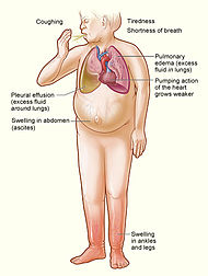
Common causes of heart failure include myocardial infarction and other forms of ischemic heart disease, hypertension, valvular heart disease and cardiomyopathy.[2] Heart failure can cause a large variety of symptoms such as shortness of breath (typically worse when lying flat, which is called orthopnea), coughing, ankle swelling and reduced exercise capacity. Heart failure is often undiagnosed due to a lack of a universally agreed definition and challenges in definitive diagnosis. Treatment commonly consist of lifestyle measures (such as decreased salt intake) and medications, and sometimes devices or even surgery.

Heart failure is a common, costly, disabling and deadly condition.[2] In developing countries, around 2% of adults suffer from heart failure, but in those over the age of 65, this increases to 6—10%.[2][3] Mostly due to costs of hospitalisation, it is associated with a high health expenditure; costs have been estimated to amount to 2% of the total budget of the National Health Service in the United Kingdom, and more than $35 billion in the United States.[4][5] Heart failure is associated with significantly reduced physical and mental health, resulting in a markedly decreased quality of life.[6][7] With the exception of heart failure caused by reversible conditions, the condition usually worsens over time. Although some patients survive many years, progressive disease is associated with an overall annual mortality rate of 10%.[8]


Terminology

Heart failure is a global term for the physiological state in which cardiac output is insufficient for the body's needs.

This may occur when the cardiac output is low (often termed "congestive heart failure").[9]

In contrast, it may also occur when the body's requirements for oxygen and nutrients are increased, and demand outstrips what the heart can provide, (termed "high output cardiac failure") [10]. This can occur in the context of severe anemia, Gram negative septicaemia, beriberi (vitamin B1/thiamine deficiency), thyrotoxicosis, Paget's disease, arteriovenous fistulae or arteriovenous malformations.

Fluid overload is a common problem for people with heart failure, but is not synonymous with it. Patients with treated heart failure will often be euvolaemic (a term for normal fluid status), or more rarely, dehydrated.

Doctors use the words "acute" to mean of rapid onset, and "chronic" of long duration. Chronic heart failure is therefore a long term situation, usually with stable treated symptomatology.

Acute decompensated heart failure, which should just describe sudden onset HF, is also used to describe exacerbated or decompensated heart failure, referring to episodes in which a patient with known chronic heart failure abruptly develops symptoms.[citation needed]

There are several terms which are closely related to heart failure, and may be the cause of heart failure, but should not be confused with it:

  • Cardiac arrest, and asystole both refer to situations in which there is no cardiac output at all. Without urgent treatment, these result in sudden death.
  • Heart attack refers to a blockage in a coronary (heart) artery resulting in heart muscle damage.
  • Cardiomyopathy refers specifically to problems within the heart muscle, and these problems usually result in heart failure. Ischemic cardiomyopathy implies that the cause of muscle damage is coronary artery disease. Dilated cardiomyopathy implies that the muscle damage has resulted in enlargement of the heart. Hypertrophic cardiomyopathy involves enlargement and thickening of the heart muscle.

[edit] Classification

There are many different ways to categorize heart failure, including:

  • the side of the heart involved, (left heart failure versus right heart failure)
  • whether the abnormality is due to contraction or relaxation of the heart (systolic dysfunction vs. diastolic dysfunction)
  • whether the problem is primarily increased venous back pressure (behind) the heart, or failure to supply adequate arterial perfusion (in front of) the heart (backward vs. forward failure)
  • whether the abnormality is due to low cardiac output with high systemic vascular resistance or high cardiac output with low vascular resistance (low-output heart failure vs. high-output heart failure)
  • the degree of functional impairment conferred by the abnormality (as in the NYHA functional classification)

Functional classification generally relies on the New York Heart Association Functional Classification.[11] The classes (I-IV) are:

  • Class I: no limitation is experienced in any activities; there are no symptoms from ordinary activities.
  • Class II: slight, mild limitation of activity; the patient is comfortable at rest or with mild exertion.
  • Class III: marked limitation of any activity; the patient is comfortable only at rest.
  • Class IV: any physical activity brings on discomfort and symptoms occur at rest.

This score documents severity of symptoms, and can be used to assess response to treatment. While its use is widespread, the NYHA score is not very reproducible and doesn't reliably predict the walking distance or exercise tolerance on formal testing.[12]

In its 2001 guidelines, the American College of Cardiology/American Heart Association working group introduced four stages of heart failure:[13]

  • Stage A: Patients at high risk for developing HF in the future but no functional or structural heart disorder;
  • Stage B: a structural heart disorder but no symptoms at any stage;
  • Stage C: previous or current symptoms of heart failure in the context of an underlying structural heart problem, but managed with medical treatment;
  • Stage D: advanced disease requiring hospital-based support, a heart transplant or palliative care.

The ACC staging system is useful in that Stage A encompasses "pre-heart failure" - a stage where intervention with treatment can presumably prevent progression to overt symptoms. ACC stage A does not have a corresponding NYHA class. ACC Stage B would correspond to NYHA Class I. ACC Stage C corresponds to NYHA Class II and III, while ACC Stage D overlaps with NYHA Class IV.

[edit] Diagnostic criteria

No system of diagnostic criteria has been agreed as the gold standard for heart failure. Commonly used systems are the "Framingham criteria"[14] (derived from the Framingham Heart Study), the "Boston criteria",[15] the "Duke criteria",[16] and (in the setting of acute myocardial infarction) the "Killip class".[17]

[edit] Signs and symptoms

[edit] Symptoms

Heart failure symptoms are traditionally and somewhat arbitrarily divided into "left" and "right" sided, recognizing that the left and right ventricles of the heart supply different portions of the circulation. However, heart failure is not exclusively backward failure (in the part of the circulation which drains to the ventricle).

There are several other exceptions to a simple left-right division of heart failure symptoms. Left sided forward failure overlaps with right sided backward failure. Additionally, the most common cause of right-sided heart failure is left-sided heart failure. The result is that patients commonly present with both sets of signs and symptoms.

[edit] Left-sided failure

Backward failure of the left ventricle causes congestion of the pulmonary vasculature, and so the symptoms are predominantly respiratory in nature. The patient will have dyspnea (shortness of breath) on exertion (dyspnée d'effort) and in severe cases, dyspnea at rest. Increasing breathlessness on lying flat, called orthopnea, occurs. It is often measured in the number of pillows required to lie comfortably, and in severe cases, the patient may resort to sleeping while sitting up. Another symptom of heart failure is paroxysmal nocturnal dyspnea, a sudden nighttime attack of severe breathlessness, usually several hours after going to sleep. Easy fatigueability and exercise intolerance are also common complaints related to respiratory compromise.

Compromise of left ventricular forward function may result in symptoms of poor systemic circulation such as dizziness, confusion and cool extremities at rest.

[edit] Right-sided failure

Backward failure of the right ventricle leads to congestion of systemic capillaries. This helps to generate excess fluid accumulation in the body. This causes swelling under the skin (termed peripheral edema or anasarca) and usually affects the dependent parts of the body first (causing foot and ankle swelling in people who are standing up, and sacral edema in people who are predominantly lying down). Nocturia (frequent nighttime urination) may occur when fluid from the legs is returned to the bloodstream while lying down at night. In progressively severe cases, ascites (fluid accumulation in the abdominal cavity causing swelling) and hepatomegaly (painful enlargement of the liver) may develop. Significant liver congestion may result in impaired liver function, and jaundice and even coagulopathy (problems of decreased blood clotting) may occur.

[edit] Signs

[edit] Left-sided failure

Common respiratory signs are tachypnea (increased rate of breathing) and increased work of breathing (non-specific signs of respiratory distress). Rales or crackles, heard initially in the lung bases, and when severe, throughout the lung fields suggest the development of pulmonary edema (fluid in the alveoli). Dullness of the lung fields to finger percussion and reduced breath sounds at the bases of the lung may suggest the development of a pleural effusion (fluid collection in between the lung and the chest wall). Cyanosis which suggests severe hypoxemia, is a late sign of extremely severe pulmonary edema.

Additional signs indicating left ventricular failure include a laterally displaced apex beat (which occurs if the heart is enlarged) and a gallop rhythm (additional heart sounds) may be heard as a marker of increased blood flow, or increased intra-cardiac pressure. Heart murmurs may indicate the presence of valvular heart disease, either as a cause (e.g. aortic stenosis) or as a result (e.g. mitral regurgitation) of the heart failure.

[edit] Right-sided failure

Physical examination can reveal pitting peripheral edema, ascites, and hepatomegaly. Jugular venous pressure is frequently assessed as a marker of fluid status, which can be accentuated by the hepatojugular reflux. If the right ventriclar pressure is increased, a parasternal heave may be present, signifying the compensatory increase in contraction strength.

[edit] Causes

[edit] Chronic Heart Failure

The predominance of causes of heart failure are difficult to analyse due to challenges in diagnosis, differences in populations, and changing prevalence of causes with age.

A 19 year study of 13000 healthy adults in the United States (the National Health and Nutrition Examination Survey (NHANES I) found the following causes ranked by Population Attributable Risk score: [18]

  1. Ischaemic Heart Disease 62%
  2. Cigarette Smoking 16%
  3. Hypertension (high blood pressure)10%
  4. Obesity 8%
  5. Diabetes 3%
  6. Valvular Heart Disease 2% (much higher in older populations)

An Italian registry of over 6200 patients with heart failure showed the following underlying causes: [19]

  1. Ischaemic Heart Disease 40%
  2. Dilated Cardiomyopathy 32%
  3. Valvular Heart Disease 12%
  4. Hypertension 11%
  5. Other 5%

Rarer causes of heart failure include:

  • Viral Myocarditis (an infection of the heart muscle)
  • Infiltrations of the muscle such as amyloidosis
  • HIV cardiomyopathy (caused by Human Immunodeficiency Virus)
  • Connective Tissue Diseases such as Systemic lupus erythematosus
  • Abuse of drugs such as alcohol
  • Pharmaceutical drugs such as chemotherapeutic agents.
  • Arrhythmias

Obstructive Sleep Apnea a condition of sleep disordered breathing overlaps with obesity, hypertension and diabetes and is regarded as an independent cause of heart failure.

[edit] Acute decompensated heart failure

See main article: Acute decompensated heart failure

Chronic stable heart failure may easily decompensate. This most commonly results from an intercurrent illness (such as pneumonia), myocardial infarction (a heart attack), arrhythmias, uncontrolled hypertension, or a patient's failure to maintain a fluid restriction, diet or medication.[20] Other well recognised precipitating factors include anaemia and hyperthyroidism which place additional strain on the heart muscle. Excessive fluid or salt intake, and medication that causes fluid retention such as NSAIDs and thiazolidinediones, may also precipitate decompensation.

Pathophysiology


Heart failure is caused by any condition which reduces the efficiency of the myocardium, or heart muscle, through damage or overloading. As such, it can be caused by as diverse an array of conditions as myocardial infarction (in which the heart muscle is starved of oxygen and dies), hypertension (which increases the force of contraction needed to pump blood) and amyloidosis (in which protein is deposited in the heart muscle, causing it to stiffen). Over time these increases in workload will produce changes to the heart itself:

  • Reduced contractility, or force of contraction, due to overloading of the ventricle. In health, increased filling of the ventricle results in increased contractility (by the Frank-Starling law of the heart) and thus a rise in cardiac output. In heart failure this mechanism fails, as the ventricle is loaded with blood to the point where heart muscle contraction becomes less efficient. This is due to reduced ability to cross-link actin and myosin filaments in over-stretched heart muscle.[22]
  • A reduced stroke volume, as a result of a failure of systole, diastole or both. Increased end systolic volume is usually caused by reduced contractility. Decreased end diastolic volume results from impaired ventricular filling – as occurs when the compliance of the ventricle falls (i.e. when the walls stiffen).
  • Reduced spare capacity. As the heart works harder to meet normal metabolic demands, the amount cardiac output can increase in times of increased oxygen demand (e.g. exercise) is reduced. This contributes to the exercise intolerance commonly seen in heart failure.
  • Increased heart rate, stimulated by increased sympathetic activity in order to maintain cardiac output. Initially, this helps compensate for heart failure by maintaining blood pressure and perfusion, but places further strain on the myocardium, increasing coronary perfusion requirements, which can lead to worsening of ischemic heart disease. Sympathetic activity may also cause potentially fatal arrhythmias.
  • Hypertrophy (an increase in physical size) of the myocardium, caused by the terminally differentiated heart muscle fibres increasing in size in an attempt to improve contractility. This may contribute to the increased stiffness and decreased ability to relax during diastole.
  • Enlargement of the ventricles, contributing to the enlargement and spherical shape of the failing heart. The increase in ventricular volume also causes a reduction in stroke volume due to mechanical and contractile inefficiency.[23]

The general effect is one of reduced cardiac output and increased strain on the heart. This increases the risk of cardiac arrest (specifically due to ventricular dysrhythmias), and reduces blood supply to the rest of the body. In chronic disease the reduced cardiac output causes a number of changes in the rest of the body, some of which are physiological compensations, some of which are part of the disease process:

  • Arterial blood pressure falls. This destimulates baroreceptors in the carotid body and aortic arch which link to the nucleus tractus solitarius. This center in the brain increases sympathetic activity, releasing catecholamines into the blood stream. Binding to alpha-1 receptors results in systemic arterial vasoconstriction. This helps restore blood pressure but also increases the total peripheral resistance, increasing the workload of the heart. Binding to beta-1 receptors in the myocardium increases the heart rate and make contractions more forceful, in an attempt to increase cardiac output. This also, however, increases the amount of work the heart has to perform.
  • Increased sympathetic stimulation also causes the hypothalamus to secrete vasopressin (also known as antidiuretic hormone or ADH), which causes fluid retention at the kidneys. This increases the blood volume and blood pressure.
  • Reduced perfusion (blood flow) to the kidneys stimulates the release of renin – an enzyme which catalyses the production of the potent vasopressor angiotensin. Angiotensin and its metabolites cause further vasocontriction, and stimulate increased secretion of the steroid aldosterone from the adrenal glands. This promotes salt and fluid retention at the kidneys, also increasing the blood volume.
  • The chronically high levels of circulating neuroendocrine hormones such as catecholamines, renin, angiotensin, and aldosterone affects the myocardium directly, causing structural remodelling of the heart over the long term. Many of these remodelling effects seem to be mediated by transforming growth factor beta (TGF-beta), which is a common downstream target of the signal transduction cascade initiated by catecholamines[24] and angiotensin II[25], and also by epidermal growth factor (EGF), which is a target of the signaling pathway activated by aldosterone[26]
  • Reduced perfusion of skeletal muscle causes atrophy of the muscle fibres. This can result in weakness, increased fatigueability and decreased peak strength - all contributing to exercise intolerance.[27]

The increased peripheral resistance and greater blood volume place further strain on the heart and accelerates the process of damage to the myocardium. Vasoconstriction and fluid retention produce an increased hydrostatic pressure in the capillaries. This shifts of the balance of forces in favour of interstitial fluid formation as the increased pressure forces additional fluid out of the blood, into the tissue. This results in edema (fluid build-up) in the tissues. In right-sided heart failure this commonly starts in the ankles where venous pressure is high due to the effects of gravity (although if the patient is bed-ridden, fluid accumulation may begin in the sacral region.) It may also occur in the abdominal cavity, where the fluid build-up is called ascites. In left-sided heart failure edema can occur in the lungs - this is called cardiogenic pulmonary oedema. This reduces spare capacity for ventilation, causes stiffening of the lungs and reduces the efficiency of gas exchange by increasing the distance between the air and the blood. The consequences of this are shortness of breath, orthopnoea and paroxysmal nocturnal dyspnea.

The symptoms of heart failure are largely determined by which side of the heart fails. The left side pumps blood into the systemic circulation, whilst the right side pumps blood into the pulmonary circulation. Whilst left-sided heart failure will reduce cardiac output to the systemic circulation, the initial symptoms often manifest due to effects on the pulmonary circulation. In systolic dysfunction, the ejection fraction is decreased, leaving an abnormally elevated volume of blood in the left ventricle. In diastolic dysfunction, end-diastolic ventricular pressure will be high. This increase in volume or pressure backs up to the left atrium and then to the pulmonary veins. Increased volume or pressure in the pulmonary veins impairs the normal drainage of the alveoli and favors the flow of fluid from the capillaries to the lung parenchyma, causing pulmonary edema. This impairs gas exchange. Thus, left-sided heart failure often presents with respiratory symptoms: shortness of breath, orthopnea and paroxysmal nocturnal dyspnea.

In severe cardiomyopathy, the effects of decreased cardiac output and poor perfusion become more apparent, and patients will manifest with cold and clammy extremities, cyanosis, claudication, generalized weakness, dizziness, and syncope

The resultant hypoxia caused by pulmonary edema causes vasoconstriction in the pulmonary circulation, which results in pulmonary hypertension. Since the right ventricle generates far lower pressures than the left ventricle (approximately 20 mmHg versus around 120 mmHg, respectively, in the healthy individual) but nonetheless generates cardiac output exactly equal to the left ventricle, this means that a small increase in pulmonary vascular resistance causes a large increase in amount of work the right ventricle must perform. However, the main mechanism by which left-sided heart failure causes right-sided heart failure is actually not well understood. Some theories invoke mechanisms that are mediated by neurohormonal activation. Mechanical effects may also contribute. As the left ventricle distends, the intraventricular septum bows into the right ventricle, decreasing the capacity of the right ventricle.

[edit] Systolic dysfunction

Heart failure caused by systolic dysfunction is more readily recognized. It can be simplistically described as failure of the pump function of the heart. It is characterized by a decreased ejection fraction (less than 45%). The strength of ventricular contraction is attenuated and inadequate for creating an adequate stroke volume, resulting in inadequate cardiac output. In general, this is caused by dysfunction or destruction of cardiac myocytes or their molecular components. In congenital diseases such as Duchenne muscular dystrophy, the molecular structure of individual myocytes is affected. Myocytes and their components can be damaged by inflammation (such as in myocarditis) or by infiltration (such as in amyloidosis). Toxins and pharmacological agents (such as ethanol, cocaine, and amphetamines) cause intracellular damage and oxidative stress. The most common mechanism of damage is ischemia causing infarction and scar formation. After myocardial infarction, dead myocytes are replaced by scar tissue, deleteriously affecting the function of the myocardium. On echocardiogram, this is manifest by abnormal or absent wall motion.

Because the ventricle is inadequately emptied, ventricular end-diastolic pressure and volumes increase. This is transmitted to the atrium. On the left side of the heart, the increased pressure is transmitted to the pulmonary vasculature, and the resultant hydrostatic pressure favors extravassation of fluid into the lung parenchyma, causing pulmonary edema. On the right side of the heart, the increased pressure is transmitted to the systemic venous circulation and systemic capillary beds, favoring extravassation of fluid into the tissues of target organs and extremities, resulting in dependent peripheral edema.

[edit] Diastolic dysfunction

Heart failure caused by diastolic dysfunction is generally described as the failure of the ventricle to adequately relax and typically denotes a stiffer ventricular wall. This causes inadequate filling of the ventricle, and therefore results in an inadequate stroke volume. The failure of ventricular relaxation also results in elevated end-diastolic pressures, and the end result is identical to the case of systolic dysfunction (pulmonary edema in left heart failure, peripheral edema in right heart failure.)

Diastolic dysfunction can be caused by processes similar to those that cause systolic dysfunction, particularly causes that affect cardiac remodeling.

Diastolic dysfunction may not manifest itself except in physiologic extremes if systolic function is preserved. The patient may be completely asymptomatic at rest. However, they are exquisitely sensitive to increases in heart rate, and sudden bouts of tachycardia (which can be caused simply by physiological responses to exertion, fever, or dehydration, or by pathological tachyarrhythmias such as atrial fibrillation with rapid ventricular response) may result in flash pulmonary edema. Adequate rate control (usually with a pharmacological agent that slows down AV conduction such as a calcium channel blocker or a beta-blocker) is therefore key to preventing decompensation.

Left ventricular diastolic function can be determined through echocardiography by measurement of various parameters such as the E/A ratio (early-to-atrial left ventricular filling ratio), the E (early left ventricular filling) deceleration time, and the isovolumic relaxation time.

[edit] Diagnosis

Chest x-ray showing an enlarged cardiac silhouette due to congestive heart failure.

[edit] Imaging

Echocardiography is commonly used to support a clinical diagnosis of heart failure. This modality uses ultrasound to determine the stroke volume (SV, the amount of blood in the heart that exits the ventricles with each beat), the end-diastolic volume (EDV, the total amount of blood at the end of diastole), and the SV in proportion to the EDV, a value known as the ejection fraction. In pediatrics, the shortening fraction is the preferred measure of systolic function. Normally, the EF should be between 50% and 70%; in systolic heart failure, it drops below 40%. Echocardiography can also identify valvular heart disease and assess the state of the pericardium (the connective tissue sac surrounding the heart). Echocardiography may also aid in deciding what treatments will help the patient, such as medication, insertion of an implantable cardioverter-defibrillator or cardiac resynchronization therapy. Echocardiography can also help determine if acute myocardial ischemia is the precipitating cause, and may manifest as regional wall motion abnormalities on echo.

Chest X-rays are frequently used to aid in the diagnosis of CHF. In the compensated patient, this may show cardiomegaly (visible enlargement of the heart), quantified as the cardiothoracic ratio (proportion of the heart size to the chest). In left ventricular failure, there may be evidence of vascular redistribution ("upper lobe blood diversion" or "cephalization"), Kerley lines, cuffing of the areas around the bronchi, and interstitial edema.

[edit] Electrophysiology

An electrocardiogram (ECG/EKG) is used to identify arrhythmias, ischemic heart disease, right and left ventricular hypertrophy, and presence of conduction delay or abnormalities (e.g. left bundle branch block). An ECG may also diagnose acute myocardial ischemia or infarction (if ST depression or elevation are present).

[edit] Blood tests

Blood tests routinely performed include electrolytes (sodium, potassium), measures of renal function, liver function tests, thyroid function tests, a complete blood count, and often C-reactive protein if infection is suspected. An elevated B-type natriuretic peptide (BNP) is a specific test indicative of heart failure. Additionally, BNP can be used to differentiate between causes of dyspnea due to heart failure from other causes of dyspnea. If myocardial infarction is suspected, various cardiac markers may be used.

According to a meta-analysis comparing BNP and N-terminal pro-BNP (NTproBNP) in the diagnosis of heart failure, BNP is a better indicator for heart failure and left ventricular systolic dysfunction. In groups of symptomatic patients, a diagnostic odds ratio of 27 for BNP compares with a sensitivity of 85% and specificity of 84% in detecting heart failure. [28]

[edit] Angiography

Heart failure may be the result of coronary artery disease, and its prognosis depends in part on the ability of the coronary arteries to supply blood to the myocardium (heart muscle). As a result, coronary catheterization may be used to identify possibilities for revascularisation through percutaneous coronary intervention or bypass surgery.

[edit] Monitoring

Various measures are often used to assess the progress of patients being treated for heart failure. These include fluid balance (calculation of fluid intake and excretion), monitoring body weight (which in the shorter term reflects fluid shifts).


Coronary disease

Coronary disease (or coronary heart disease) refers to the failure of coronary circulation to supply adequate circulation to cardiac muscle and surrounding tissue.

It is sometimes equated with atherosclerotic coronary artery disease, but coronary disease can be due to other causes, such as coronary vasospasm.[1] It is possible for the stenosis to be caused by the spasm.

Left ventricular hypertrophy

Left ventricular hypertrophy (LVH) is the thickening of the myocardium (muscle) of the left ventricle of the heart.

Causes

While ventricular hypertrophy occurs naturally as a reaction to aerobic exercise and strength training, it is most frequently referred to as a pathological reaction to cardiovascular disease, or high blood pressure.

While LVH itself is not a disease, it is usually a marker for disease involving the heart.[2] Disease processes that can cause LVH include any disease that increases the afterload that the heart has to contract against, and some primary diseases of the muscle of the heart.

Causes of increased afterload that can cause LVH include aortic stenosis, aortic insufficiency, and hypertension. Primary disease of the muscle of the heart that cause LVH are known as hypertrophic cardiomyopathies, which can lead into heart failure.

Diagnosis

The principal method to diagnose LVH is echocardiography, during which the thickness of the muscle of the heart can be measured. The electrocardiogram (ECG) often shows signs of increased voltage from the heart in individuals with LVH, so this is often used as a screening test to determine who should undergo further testing with an echocardiogram.

Echocardiography

Two dimensional echocardiography can produce images of the left ventricle. The thickness of the left ventricle as visualized on echocardiography correlates with its actual mass. Normal thickness of the left ventricular myocardium is from 0.6 to 1.1 cm (as measured at the very end of diastole. If the myocardium is more than 1.1 cm thick, the diagnosis of LVH can be made.

ECG criteria for LVH

There are several sets of criteria used to diagnose LVH via electrocardiography.[3] None of them are perfect, though by using considering multiple criteria sets, the sensitivity and specificity are increased.

The Sokolow-Lyon index:

  • S in V1 + R in V5 or V6 (whichever is larger) ≥ 35 mm
  • R in aVL ≥ 11 mm

The Cornell voltage criteria for the ECG diagnosis of LVH involves measurement of the sum of the R wave in lead aVL and the S wave in lead V3. The Cornell criteria for LVH are:

  • S in V3 + R in aVL > 28 mm (men)
  • S in V3 + R in aVL > 20 mm (women)

Romhilt-Estes point score system ("diagnostic" >5 points; "probable" 4 points):

ECG Criteria Points
Voltage Criteria (any of):
  1. R or S in limb leads ≥20 mm
  2. S in V1 or V2 ≥30 mm
  3. R in V5 or V6 ≥30 mm
3
ST-T Abnormalities:
  • ST-T vector opposite to QRS without digitalis
  • ST-T vector opposite to QRS with digitalis

3
1

Negative terminal P mode in V1 1 mm in depth and 0.04 sec in duration (indicates left atrial enlargement) 3
Left axis deviation (QRS of -30° or more) 2
QRS duration ≥0.09 sec 1
Delayed intrinsicoid deflection in V5 or V6 (>0.05 sec) 1

Other voltage-based criteria for LVH include:

  • Lead I: R wave > 14 mm
  • Lead aVR: S wave > 15 mm
  • Lead aVL: R wave > 12 mm
  • Lead aVF: R wave > 21 mm
  • Lead V5: R wave > 26 mm
  • Lead V6: R wave > 20 mm

Heart disease


http://www.dtsg.com/sitebuilder/images/banner-779x251.jpg

Heart disease
is an umbrella term for a variety for different diseases affecting the heart. As of 2007, it is the leading cause of death in the United States,[1][2] England, Canada and Wales,[3] killing one person every 34 seconds in the United States alone.

Types of heart disease

Coronary heart disease

Coronary artery disease is a disease of the artery caused by the accumulation of atheromatous plaques within the walls of the arteries that supply the myocardium. Angina pectoris (chest pain) and myocardial infarction (heart attack) are symptoms of and conditions caused by coronary heart disease.

Over 459,000 Americans die of coronary heart disease every year[5]. In the United Kingdom, 101,000 deaths annually are due to coronary heart disease.[6]

Cardiomyopathy

Cardiomyopathy literally means "heart muscle disease" (Myo= muscle, pathy= disease) It is the deterioration of the function of the myocardium (i.e., the actual heart muscle) for any reason. People with cardiomyopathy are often at risk of arrhythmia and/or sudden cardiac death.

  • Extrinsic cardiomyopathies - cardiomyopathies where the primary pathology is outside the myocardium itself. Most cardiomyopathies are extrinsic, because by far the most common cause of a cardiomyopathy is ischemia. The World Health Organization calls these specific cardiomyopathies[citation needed]:
    • Alcoholic cardiomyopathy
    • Coronary artery disease
    • Congenital heart disease - see below
    • Nutritional diseases affecting the heart
    • Ischemic (or ischaemic) cardiomyopathy
    • Hypertensive cardiomyopathy
    • Valvular cardiomyopathy - see also Valvular heart disease below
    • Inflammatory cardiomyopathy - see also Inflammatory heart disease below
    • Cardiomyopathy secondary to a systemic metabolic disease
  • Intrinsic cardiomyopathies - weakness in the muscle of the heart that is not due to an identifiable external cause.
    • Dilated cardiomyopathy (DCM) - most common form, and one of the leading indications for heart transplantation. In DCM the heart (especially the left ventricle) is enlarged and the pumping function is diminished.
    • Hypertrophic cardiomyopathy (HCM or HOCM) - genetic disorder caused by various mutations in genes encoding sarcomeric proteins. In HCM the heart muscle is thickened, which can obstruct blood flow and prevent the heart from functioning properly.
    • Arrhythmogenic right ventricular cardiomyopathy (ARVC) - arises from an electrical disturbance of the heart in which heart muscle is replaced by fibrous scar tissue. The right ventricle is generally most affected.
    • Restrictive cardiomyopathy (RCM) - least common cardiomyopathy. The walls of the ventricles are stiff, but may not be thickened, and resist the normal filling of the heart with blood. ** Noncompaction Cardiomyopathy - the left ventricle wall has failed to properly grow from birth and such has a spongy appearance when viewed during an echocardiogram.

http://www.nhlbi.nih.gov/health/dci/images/heart_coronary_artery.gif

Cardiovascular disease

Cardiovascular disease is any of a number of specific diseases that affect the heart itself and/or the blood vessel system, especially the veins and arteries leading to and from the heart. Research on disease dimorphism suggests that women who suffer with cardiovascular disease usually suffer from forms that affect the blood vessels while men usually suffer from forms that affect the heart muscle itself. Known or associated causes of cardiovascular disease include diabetes mellitus, hypertension, hyperhomocysteinemia and hypercholesterolemia.

Types of cardiovascular disease include:

  • Atherosclerosis

Ischaemic heart disease

  • Ischaemic heart disease - another disease of the heart itself, characterized by reduced blood supply to the organs.

chd is when the arteries that supply the oxygen and the nutrients gets stopped and the heart will not get enough of the oxygen and the nutrients and will eventually stop beating.

Heart failure

Heart failure, also called congestive heart failure (or CHF), and congestive cardiac failure (CCF), is a condition that can result from any structural or functional cardiac disorder that impairs the ability of the heart to fill with or pump a sufficient amount of blood throughout the body.

  • Cor pulmonale, a failure of the right side of the heart.

Hypertensive heart disease

Hypertensive heart disease is heart disease caused by high blood pressure, especially localised high blood pressure. Conditions that can be caused by hypertensive heart disease include:

Inflammatory heart disease

Inflammatory heart disease involves inflammation of the heart muscle and/or the tissue surrounding it.

  • Endocarditis - inflammation of the inner layer of the heart, the endocardium. The most common structures involved are the heart valves.
  • Inflammatory cardiomegaly
  • Myocarditis - inflammation of the myocardium, the muscular part of the heart.

Valvular heart disease

Valvular heart disease is disease process that affects one or more valves of the heart. The valves in the right side of the heart are the tricuspid valve and the pulmonic valve. The valves in the left side of the heart are the mitral valve and the aortic valve.

  • Aortic valve stenosis
  • Mitral valve prolapse
  • Valvular cardiomyopathy
http://www.elslaw.com/images/diagram_chronic_severe_aortic.gif